

Interactive CT & MRI Anat.Lite

Interactive CT & MRI Anat.Lite açıklaması
★Lite version★
This is the free Lite version of "Interactive CT and MRI Anatomy".
The function is restricted.
You can only see the transverse CT images of the head.
Please check the operation before purchasing the full version.
★ Details ★
This application is developed for medical students, interns, residents, doctors, nurses, and radiology technicians to understand the essential anatomical terms of the body.
You can learn anatomy by answering the terms by step-to-step questions using a total of 241 CT and MRI images.
A total of 17 images of 3D-CT, MRA and plain X-ray film(particularly the extremities) are included as references.
Other reference images include coronary artery segments defined by the American Heart Association(AHA), pulmonary segments, and liver segments(according to Couinaud classification).
You can enlarge all the images by simple manipulation.
★ Major functions ★
There are 4 major functions.
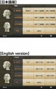
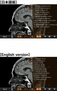
-1) Anatomical mode
Anatomical terms are overlaid on the images.
It can be used as the anatomical atlas.
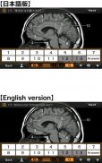
-2) Quiz mode type 1
You select the part of the image by using anatomical term.
Questions will basically appear randomly.
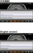
-3) Quiz mode type 2
You select the anatomical term by the part of the image.
Questions will basically appear randomly.
-4) Index
You can find the specific images by using anatomical terms.
★ Intended users ★
-Medical students
-Interns and residents
-Doctrors
-Nurses
-Radiology technicians
-All those who are intrested in CT and MRI anatomy
★ Images(a total of 258 images) ★
Images basically include horizontal, coronal, and sagital planes.
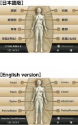
-Head(36 images including CTA and 3D-CT)
-Neck(24 images)
-Spine(19 images including plain X-ray films)
-Chest(61 images including 3D-CT images)
-Abdomen (37 images)
-Pelves: male (9 images)
-Pelvis: female (11 images)
-Extremities (shoulder, hand, elbow, hip joint, knee, foot) (61 images including plain X-ray films)
Editors
Toshiaki Nitori, M.D. (Professor of Radiology, Kyorin University, School of Medicine)
Yasuo Sasaki, M.D. (Manager of diagnostic radiology, Iwate Prefectural Central Hospital)
</div> <div jsname="WJz9Hc" style="display:none">★ Lite sürümü ★
Bu "Etkileşimli BT ve MRG Anatomy" ücretsiz Lite sürümü.
fonksiyonu sınırlıdır.
Sadece kafanın enine BT görüntülerini görebilirsiniz.
Tam sürümünü satın almadan önce çalışmasını kontrol edin.
★ ★ Detaylar
Bu uygulama vücudun temel anatomik terimleri anlamak için tıp öğrencileri, stajyerler, sakinleri, doktor, hemşire ve radyoloji teknisyenleri için geliştirilmiştir.
Sen 241 BT ve MRG görüntüleri toplam kullanarak adım için adım sorular terimleri cevaplayarak anatomi öğrenebilirsiniz.
3B-BT, MRA ve düz röntgen filminde (özellikle ekstremite) 17 görüntülerin toplam referanslar olarak dahil edilmiştir.
Diğer referans görüntüleri (Couinaud sınıflandırmasına göre) Amerikan Kalp Derneği (AHA) tarafından tanımlanan koroner arter segmentleri, akciğer segmentleri ve karaciğer segmentlerini içerir.
Basit manipülasyonla tüm görüntüleri büyütebilirsiniz.
★ Önemli fonksiyonlar ★
4 önemli işlevleri vardır.
-1) Anatomik modu
Anatomik terimler görüntülerde üst üste.
Anatomik atlas olarak kullanılabilir.
-2) Sınav modu tip 1
Sen anatomik terim kullanarak görüntünün bir bölümünü seçin.
Sorular temelde rastgele görünecektir.
-3) Sınav modu tip 2
Görüntünün bir parçası anatomik terim seçin.
Sorular temelde rastgele görünecektir.
-4) Endeksi
Sen anatomik terimleri kullanarak belirli görüntüleri bulabilirsiniz.
★ Amaçlanan kullanıcılar ★
-Medikal Öğrenciler
-Interns Ve sakinleri
-Doctrors
-Nurses
-Radiology Teknisyenleri
BT ve MRG anatomisi intrested olanlar -Tüm
★ Görüntüler (258 görüntülerin bir toplam) ★
Görüntüler temelde yatay koronal, sagital ve uçakları yer alıyor.
-Baş (CTA ve 3B-BT dahil 36 fotoğraf)
Boyun (24 fotoğraf)
-Spine (Düz röntgen filmleri de dahil olmak üzere 19 fotoğraf)
-Göğüs (3B-BT görüntüler dahil olmak üzere 61 fotoğraf)
-Abdomen (37 fotoğraf)
-Pelves: Erkek (9 fotoğraf)
-Pelvis: Kadın (11 fotoğraf)
-Extremities (Omuz, el, dirsek, kalça eklemi, diz, ayak) (düz röntgen filmleri de dahil olmak üzere 61 fotoğraf)
Editörler
Toshiaki Nitori, MD (Radyoloji Profesörü Kyorin Üniversitesi Tıp Fakültesi)
Yasuo Sasaki, MD (diagnostik radyoloji Müdürü, Iwate Valiliği Merkez Hastane)</div> <div class="show-more-end">


